45 external structure of the heart with labels
The 3 Layers of the Heart Wall - ThoughtCo The heart is an extraordinary organ. It is about the size of a clenched fist, weighs about 10.5 ounces and is shaped like a cone. Along with the circulatory system, the heart works to supply blood and oxygen to all parts of the body. The heart is located in the chest cavity just posterior to the breastbone, between the lungs, and superior to the diaphragm. How to Draw the Internal Structure of the Heart (with Pictures) To draw the internal structure of a human heart, follow the steps below. Part 1 Finding a Diagram 1 To find a good diagram, go to Google Images, and type in "The Internal Structure of the Human Heart". Find an image that displays the entire heart, and click on it to enlarge it. 2 Find a piece of paper and something to draw with.
› articles › flat-vs-deep-hierarchyFlat vs. Deep Website Hierarchies - Nielsen Norman Group Nov 10, 2013 · Left: a flat site hierarchy, with few vertical levels. Right: a deep site hierarchy has the same information organized into more sublevels. Both of these site hierarchies start at the top with a single homepage, but the information below that page is organized quite differently: the website on the left has 8 major categories, but the site on the right has only 4.

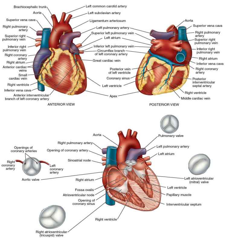
External structure of the heart with labels
Human Heart - Diagram and Anatomy of the Heart - Innerbody Because the heart points to the left, about 2/3 of the heart's mass is found on the left side of the body and the other 1/3 is on the right. Anatomy of the Heart Pericardium. The heart sits within a fluid-filled cavity called the pericardial cavity. The walls and lining of the pericardial cavity are a special membrane known as the pericardium. The Anatomy of the Heart, Its Structures, and Functions The heart is the organ that helps supply blood and oxygen to all parts of the body. It is divided by a partition (or septum) into two halves. The halves are, in turn, divided into four chambers. The heart is situated within the chest cavity and surrounded by a fluid-filled sac called the pericardium. This amazing muscle produces electrical ... Heart Anatomy: size, location, coverings and layers : Anatomy & Physiology The heart wall is composed of three layers: the epicardium, myocardium, and endocardium. Location of the heart in the mediastinum. The superficial epicardium is the visceral layer of the serous pericardium. The middle layer is the myocardium and is composed mainly of cardiac muscle and forms the bulk of the heart.
External structure of the heart with labels. Diagrams, quizzes and worksheets of the heart - Kenhub Worksheet showing unlabelled heart diagrams. Using our unlabeled heart diagrams, you can challenge yourself to identify the individual parts of the heart as indicated by the arrows and fill-in-the-blank spaces. This exercise will help you to identify your weak spots, so you'll know which heart structures you need to spend more time studying ... study.com › learn › lessonWhat is Trachea? | Function, Structure and Purpose - Video ... Jul 02, 2021 · This structure is a large, thick piece of cartilage that covers the larynx and superior opening of the trachea to provide protection for these delicate structures. The larynx and epiglottis make ... Solved Help Label the external anatomy on this posterior - Chegg Question: Help Label the external anatomy on this posterior view of a mammalian heart by clicking and dragging the labels to the correct location Coronary sinus Apex of heart Lert atrium Posterior interventricular branch of LCA Left pulmonary artery Left ventricle Left pulmonary veins Aortic arch This problem has been solved! See the answer A Labeled Diagram of the Human Heart You Really Need to See The human heart, comprises four chambers: right atrium, left atrium, right ventricle and left ventricle. The two upper chambers are called the left and the right atria, and the two lower chambers are known as the left and the right ventricles. The two atria and ventricles are separated from each other by a muscle wall called 'septum'.
quizlet.com › 574029087 › ch-19-circulatory-systemCh. 19 Circulatory System- heart Flashcards | Quizlet Correctly label the external anatomy of the anterior heart. Place the labels in order denoting the flow of blood through the pulmonary circuit beginning with the right atrium and ending in the left atrioventricular valve. The first and last structures are given. Right atrium 1. tricuspid valve 2. right ventricle 3. pulmonary valve byjus.com › biology › human-heartHuman Heart - Anatomy, Functions and Facts about Heart The external structure of the heart has many blood vessels that form a network, with other major vessels emerging from within the structure. The blood vessels typically comprise the following: Veins supply deoxygenated blood to the heart via inferior and superior vena cava, and it eventually drains into the right atrium. Structure Of The Heart | A-Level Biology Revision Notes The two ventricles: these are the lower two chambers. They have thick, muscular walls which pump blood through the arteries. The heart is divided longitudinally into two sides by means of muscular walls. Each atrium is connected to its own ventricle through an opening which is guarded by a valve. The Heart - Science Quiz - GeoGuessr This science quiz game will help you identify the parts of the human heart with ease. Blood comes in through veins and exists via arteries—to control the direction of the flow, the heart has four sets of valves. The heart is an amazing machine with a lot of moving parts—let this quiz game help you find your way around this most vital of organs.
Chapter 22 Heart Flashcards - Quizlet Label the coronary arteries in an anterior view of the heart. Label the order that blood flows through in the heart, using the arrows as guides. Label the components of the heart wall. Label the components of the heart as seen from a posterior view. Label the major coronary veins. Label the components of the conduction system. Correctly Label The Following External Anatomy Of The Anterior Heart ... The right atrium, the anterior descending aorta, and the vasculature are all essential organs of the cardiovascular system. The external anatomy of the human heart consists of the four chambers that form the apex of the heart. Each chamber has an apex that corresponds to a box. There are two boxes on each side of the heart: the atria and the ... Structure of the Heart | SEER Training The human heart is a four-chambered muscular organ, shaped and sized roughly like a man's closed fist with two-thirds of the mass to the left of midline. The heart is enclosed in a pericardial sac that is lined with the parietal layers of a serous membrane. The visceral layer of the serous membrane forms the epicardium. Layers of the Heart Wall en.wikipedia.org › wiki › DapagliflozinDapagliflozin - Wikipedia Medical uses. Dapagliflozin is used along with diet, exercise and usually with other glucose lowering medications, to improve glycaemic control in adults with type 2 diabetes and to reduce the risk of hospitalisation for heart failure among adults with type 2 diabetes and known cardiovascular disease or other cardiovascular risk factors (including high blood pressure, high cholesterol and ...
Comparative Anatomy Tutorial - External Anatomy
Heart Anatomy Labeling Game - PurposeGames.com This is an online quiz called Heart Anatomy Labeling Game There is a printable worksheet available for download here so you can take the quiz with pen and paper. Your Skills & Rank Total Points 0 Get started! Today's Rank -- 0 Today 's Points One of us! Game Points 19 You need to get 100% to score the 19 points available Actions

The Structure & Functions of the Heart - Elite Cardiovascular Group
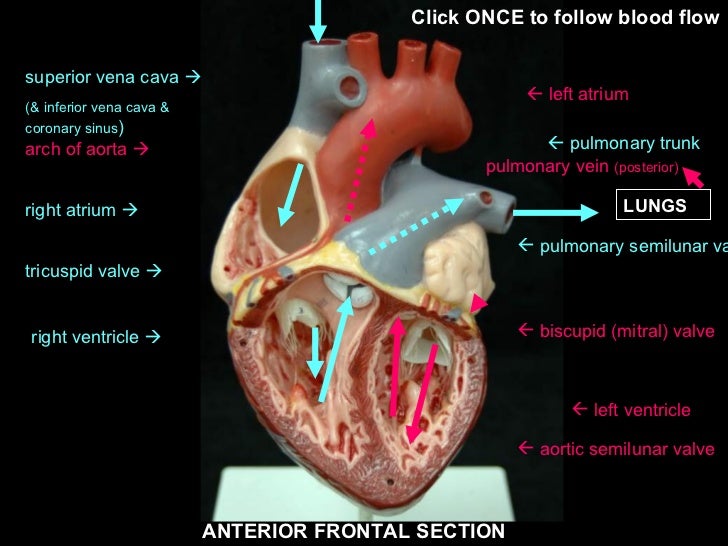
Label the HEART | Circulatory System Quiz - Quizizz True or False: Blood flows in the following sequence in the heart: Vena cava, right atrium, right ventricle, pulmonary artery, lungs, pulmonary veins, left atrium, left ventricle, aorta. Q. True or False: There are four chambers in the heart. Q. Place the pathway of blood through the heart in the correct sequence. Q.
Heart without labels
External anterior heart labeling Quiz - PurposeGames.com About this Quiz This is an online quiz called External anterior heart labeling There is a printable worksheet available for download here so you can take the quiz with pen and paper. Your Skills & Rank Total Points 0 Get started! Today's Rank -- 0 Today 's Points One of us! Game Points 27 You need to get 100% to score the 27 points available

The Heart - Biology Student
Heart Diagram with Labels and Detailed Explanation - BYJUS Diagram of Heart. The human heart is the most crucial organ of the human body. It pumps blood from the heart to different parts of the body and back to the heart. The most common heart attack symptoms or warning signs are chest pain, breathlessness, nausea, sweating etc. The diagram of heart is beneficial for Class 10 and 12 and is frequently ...

Basics about Cardiovascular System : Structure and Function
Heart Anatomy | Anatomy and Physiology | | Course Hero Learning Objectives. By the end of this section, you will be able to: Describe the location and position of the heart within the body cavity. Describe the internal and external anatomy of the heart. Identify the tissue layers of the heart. Relate the structure of the heart to its function as a pump. Compare systemic circulation to pulmonary ...

mypicsainmarin: heart diagram with labels
Lesson | The Heart - External Structure | Encounter Edu In this lesson students begin their exploration of the circulatory system, labelling a diagram of the external structures and identifying arteries and veins. They will go on to explain where blood enters and leaves the heart. Learning outcomes
Anatomy Review: The Heart
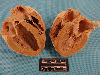
pmt.physicsandmathstutor.com › download › BiologyPractical notes - SP 2.3c Dissection of a Mammalian Heart ... The mammalian heart is a muscular pump that pushes blood around the body. It consists of four chambers and associated blood vessels . The left and right side of the heart is separated by a muscular wall, the septum . Recall the structure of the heart in the diagram below:
heart
quizlet.com › 630625176 › chapter-19-the-heart-flashChapter 19: The Heart Flashcards | Quizlet •Allows heart to beat without friction, gives it room to expand and resists excessive expansion •Parietal pericardium-tough outer, fibrous layer of connective tissue-inner serous layer •Visceral pericardium (a.k.a. epicardium of heart wall)-serous lining of sac turns inward at base of heart to cover the heart surface

Pin on Classical Conversations Science
Labelling the heart — Science Learning Hub Blood transports oxygen and nutrients to the body. It is also involved in the removal of metabolic wastes. In this interactive, you can label parts of the human heart. Drag and drop the text labels onto the boxes next to the diagram. Selecting or hovering over a box will highlight each area in the diagram.

The Cardiovascular System: Anatomy & Physiology - The Nursing Journal
Solved -labeling Activity: External Anatomy of the Sheep | Chegg.com Anatomy and Physiology. Anatomy and Physiology questions and answers. -labeling Activity: External Anatomy of the Sheep Heart Part A Drag the labels to the appropriate location in the figure. Reset Help Lolt ventric Pulmonary trunk Lolt atrium Lohtaude Right trum Posterior Interventricular sules = Pulmonary veins Art Right vorticle Anterior ...

External Structure Of Heart Anatomy Diagram | MedicineBTG.com
Heart - External Features - Anatomy QA Apex beat. Is the lowermost and outermost thrust of the heart, felt on the front of the chest. In adults it is felt in the left 5 th intercostal space 9cm. from the median plane (just medial to the midclavicular line). In infants it is felt in the 3 rd intercostal space just lateral to the midclavicular line.. Dextrocardia. It is a congenital anomaly in which the heart lies on the right side ...

The Heart | S-cool, the revision website
The structure of the heart - Structure and function of the heart ... Each side of the heart consists of an atrium and a ventricle which are two connected chambers. The atria (plural of atrium) are where the blood collects when it enters the heart. The ventricles...
iGCSE Biology - Gross Structure Of The Heart - BioChem Tuition
Layers of the heart: Epicardium, myocardium, endocardium - Kenhub The endocardium is the innermost layer of the heart. It lines the inner surfaces of the heart chambers, including the heart valves. The endocardium has two layers. The inner layer lines the heart chambers and is made of endothelial cells.

31 Blood Vessels Diagram To Label
Heart Anatomy: Heart Dissection - University of Washington The major vessels of the heart are found at the base of the heart, along with the upper chambers, the right atrium (C) and left atrium (D). The atria are collapsed, but in a functioning heart, they would be stretched full of blood. The majority of the heart tissue consists of the ventricles. The left ventricle (F) is stiff and solid because it ...

Aqua Fanatic: Crayfish Anatomy
Heart Labeling Quiz: How Much You Know About Heart Labeling? Here is a Heart labeling quiz for you. The human heart is a vital organ for every human. The more healthy your heart is, the longer the chances you have of surviving, so you better take care of it. Take the following quiz to know how much you know about your heart. Questions and Answers. 1.
Post a Comment for "45 external structure of the heart with labels"